Contents
- 1 The unceasing inventions and iterations in dental applied sciences and fabrics have taken the sophistication of medicine modalities to a historical stage. On the other hand, once we are centered at the newness, the fun of the end result, we fail to be aware of the aim of the job. A working example is dental implants. Those have been first of all thought to be the gold same old for compromised teeth alternative in spite of reviews that endodontic remedies ensured capability over the years to be within the vary of 91%–97%.1 The novelty was tarnished by means of reviews that peri-implantitis and peri-mucositis confirmed an average weighted occurrence price of 43% throughout Europe and 22% throughout South and North The united states.2 A myriad of things influences the initiation and development of peri-implant illness; sadly, the medicine of peri-implant illness is at absolute best beneficial within the quick time period, the illness having a prime price of continual irritation and recurrence.3 A find out about by means of Guarnieri et al. confirmed that energetic periodontal treatment in continual periodontal sufferers adopted by means of long-term common periodontal repairs used to be a success within the retention of nearly all of periodontally compromised tooth.4 In the similar sufferers, a better tendency for implant loss than teeth loss used to be discovered. The sequence of case reviews introduced in this article is going to supply choice information classes for absolute best practices which facilitate retention of compromised tooth the usage of endodontic remedies. Case record 1: CBCT in endodontics
- 2 Case record 2: Root amputation (periodontal and endodontic lesion)
- 3 Case record 3: Resorptive defect
- 4 Case record 4: Cracked teeth
- 5 Conclusion
The unceasing inventions and iterations in dental applied sciences and fabrics have taken the sophistication of medicine modalities to a historical stage. On the other hand, once we are centered at the newness, the fun of the end result, we fail to be aware of the aim of the job. A working example is dental implants. Those have been first of all thought to be the gold same old for compromised teeth alternative in spite of reviews that endodontic remedies ensured capability over the years to be within the vary of 91%–97%.1 The novelty was tarnished by means of reviews that peri-implantitis and peri-mucositis confirmed an average weighted occurrence price of 43% throughout Europe and 22% throughout South and North The united states.2
A myriad of things influences the initiation and development of peri-implant illness; sadly, the medicine of peri-implant illness is at absolute best beneficial within the quick time period, the illness having a prime price of continual irritation and recurrence.3 A find out about by means of Guarnieri et al. confirmed that energetic periodontal treatment in continual periodontal sufferers adopted by means of long-term common periodontal repairs used to be a success within the retention of nearly all of periodontally compromised tooth.4 In the similar sufferers, a better tendency for implant loss than teeth loss used to be discovered. The sequence of case reviews introduced in this article is going to supply choice information classes for absolute best practices which facilitate retention of compromised tooth the usage of endodontic remedies.
Case record 1: CBCT in endodontics
Most likely an important good thing about CBCT in endodontics is that it demonstrates anatomical options in three-D that intra-oral, panoramic and cephalometric pictures can’t. As well as, as a result of reconstruction of CBCT knowledge is carried out natively the usage of a non-public laptop, knowledge can also be reoriented in true spatial relationships.5
In October 2015, a 55-year-old male affected person, up to now observed for medicine within the place of job, self-referred for a 2d opinion in regards to the really helpful extraction of a suspected fractured teeth within the maxillary proper quadrant. He reported {that a} tender swelling had grow to be obvious over the last week. Medical exam famous fluctuant swelling between teeth #15 and teeth #14, and a 12 mm probing defect used to be detected alongside the mesiobuccal line attitude of teeth #15.
The affected person equipped a periapical radiograph appearing a mesio-proximal periradicular radiolucency related to teeth #15 (Fig. 1). The sagittal slice of the CBCT quantity (Carestream CS 9000, Carestream Dental) confirmed the level of the lesion (Fig. 2). Within the earlier root canal treatment, handiest the buccal canal have been detected and handled. The axial slice confirmed an untreated palatal canal (Fig. 3). A fibre publish positioned within the buccal canal have been used to retain the core. The affected person used to be urged of the misdirected anchoring pin extending into the periodontal ligament. With the affected person’s consent, it used to be determined to selectively deal with the palatal canal.
After an intervening time six-week length of calcium hydroxide treatment (UltraCal XS, Ultradent; Figs. 4 & 5),6 the basis canal house used to be obturated the usage of a heat vertical condensation method.7 The obturation subject matter used to be expressed right into a lateral branching portal of go out (Fig. 6). The four-year follow-up confirmed solution of the lateral lesion (Figs. 7–9). The preliminary presumption of a fractured root used to be confirmed false, suggesting that diagnosing prerequisites in line with inadequate knowledge acquisition is unreliable. Using CBCT is an crucial in endodontic procedures of any type equipped ALARA (as little as relatively achievable) ideas relating to radiation dose are adopted.
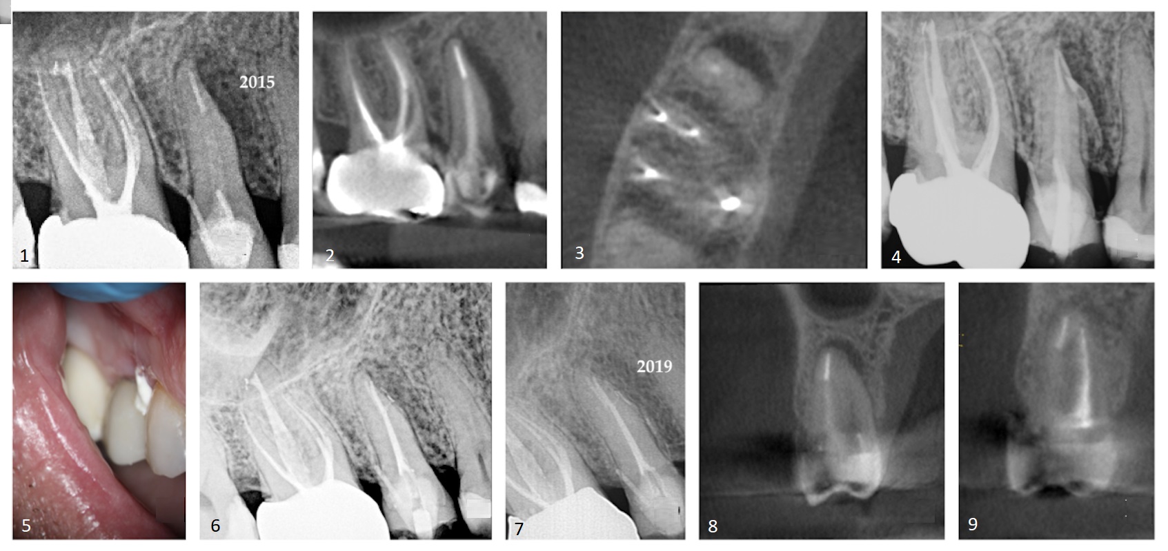
Case 1—Fig. 1: A space of periradicular rarefaction used to be glaring alongside the mesio-proximal side of teeth #15. Earlier root canal treatment and a pin-retained publish and core supporting a zirconia crown have been famous. Fig. 2: The sagittal slice of the CBCT quantity confirmed the lateral lesion extending to the alveolar crest. Fig. 3: The axial slice of the CBCT quantity confirmed the level of the rarefaction adjoining to the mesial side of the basis and the presence of an untreated palatal canal. Fig. 4: Selective medicine of the palatal canal used to be carried out. Calcium hydroxide used to be inserted within the canal house. Fig. 5: The extrusion of the intervening time calcium hydroxide medicament in the course of the sulcular space of teeth #15 used to be glaring. Fig. 6: A lateral department of the basis canal house containing the obturation subject matter exited into the interface of the center and apical thirds of the basis. Fig. 7: A periapical radiograph taken 4 years after medicine confirmed osseous regeneration and the reformation of the periodontal ligament. Fig. 8: The coronal slice of the CBCT quantity confirmed the pre-op periradicular radiolucency. Fig. 9: The coronal slice of the CBCT quantity taken 4 years after medicine confirmed the solution of the periradicular radiolucency.
Case record 2: Root amputation (periodontal and endodontic lesion)
Root resection is a medicine choice for molars with complex furcation involvement. In a find out about by means of Derks et al., mandibular molars after root resection confirmed a survival chance of virtually 80% even 20 years later.8
In October 1998, a 39-year-old male affected person introduced to the place of job complaining of gingival tenderness within the mandibular proper quadrant. In depth bone loss used to be famous interproximally between teeth #47 and teeth #46; alternatively, the periodontal standing of the dentition on the whole used to be inside commonplace limits. Pulp sensibility checking out of the tooth within the mandibular proper quadrant known the pulp of teeth #46 to be necrotic (Fig. 10). It used to be defined to the affected person that medicine of periodontal and endodontic lesions used to be on the whole destructive and that good fortune depended at the severity of bone loss, root trunk duration, stage of root separation, curvature of the basis to be resected, talent to remove the osseous defect, pulpal standing, and restorative and oral hygiene procedures required.
With the affected person’s consent, the distal root of teeth #46 used to be resected and the overlying crown portion retained (Fig. 11). Someday after the amputation process, the referring dentist splinted tooth #47 and 46 with a composite and Ribbond bridge. Twenty-two years after the preliminary process, osseous regeneration and cortication within the furcal area have been glaring between tooth #47 and 46 (Fig. 12).
Developments in methods to care for compromised tooth in live performance with a better figuring out of chance components related to dental implants invite a reassessment of the advantages of strategic extraction of a teeth with a questionable diagnosis or of restricted strategic price.9 With using hard- and soft-tissue augmentation procedures, platelet-rich fibrin, minimally invasive flap design and suturing tactics along side surgical running microscopes, it’s unreasonable to sacrifice a teeth for an implant when this venerable medicine choice displays beneficial diagnosis and good fortune charges.10
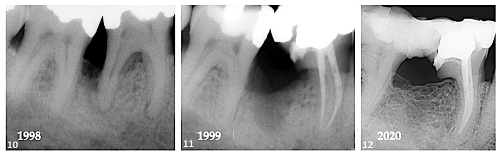
Case 2—Fig. 10: Lack of bone between teeth #47 and teeth #46 and lack of the periodontal ligament across the apical area of the mesial and distal roots have been glaring. Fig. 11: After resection of the distal root, the stage of bone loss seemed to have greater. Fig. 12: A 22-year follow-up confirmed regeneration of the misplaced interproximal bone and cortication of the alveolar crest.
Case record 3: Resorptive defect
Innovative interior resorption or the ones circumstances with perforations of the basis can also be outstanding from exterior resorption by means of various radiographic tactics. In tooth with interior resorption, the radiolucent lesion strikes with the canal when the radiographs are taken at other angles, while in exterior resorption the radiolucent lesion strikes out of doors of the canal.11, 12
In March 1999, a 47-year-old feminine affected person introduced to the place of job with facial swelling, and teeth #11 used to be mushy to percussion. A periapical radiograph of the maxillary anterior sextant confirmed huge Elegance III and IV restorations. Teeth #11 exhibited periapical rarefaction. Teeth #21 confirmed interior resorption on the mid-root stage (Fig. 13). The affected person reported that teeth #21 have been traumatised some 35 years sooner than. Within the absence of CBCT imaging, it used to be assumed that the resorption had perforated the facial side of the basis and disrupted the overlying cortical bone.
Pulpal sensibility checking out of the tooth within the anterior decided that teeth #11 used to be pulpless. The teeth used to be endodontically handled and obturated the usage of a heat vertical condensation method. Teeth #21 used to be instrumented to the incisal stage of the resorptive defect, and the canal sealed with white mineral trioxide combination (Fig. 14).13 Had been the teeth to be handled lately, the selection of obturation subject matter could be EndoSequence BC putty (Brasseler). Recently, there’s a pattern against using an injectable platelet-rich fibrin regenerative way to unravel interior resorptive defects that displays nice attainable for long-term therapeutic.14 Apply-up after 21 years confirmed that the periradicular radiolucency related to teeth #11 had resolved and that the resorptive defect have been contracted (Figs. 15 & 16).
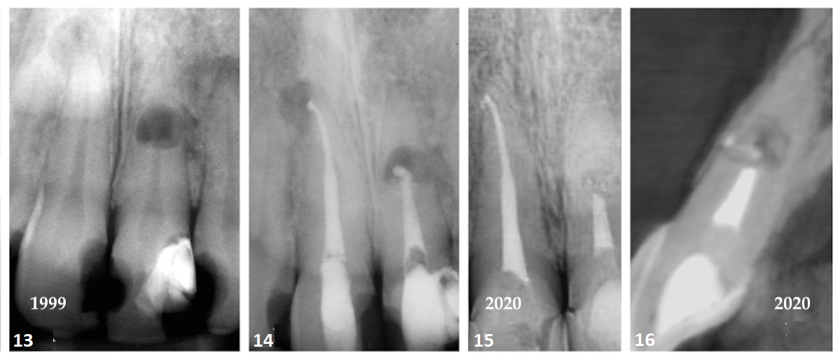
Case 3—Fig. 13: The intra-oral periapical radiograph printed a periradicular radiolucency on the root apex of teeth #11. A big space of interior resorption used to be glaring mid-root of teeth #21. The resorption had perforated the lateral side of the basis, inflicting disruption of the interproximal bone. Fig. 14: A post-op radiograph confirmed the endodontic medicine of teeth #11. Teeth #21 used to be sealed with white mineral trioxide combination to the incisal stage of the resorptive defect, because it demonstrated minimum root discoloration. Fig. 15: The periradicular radiolucency related to teeth #11 had resolved. The resorptive defect used to be diminished, and radiolucent deposits have been glaring inside the resorptive crypt. Fig. 16: The sagittal slice of the CBCT quantity confirmed an intact cortical plate. The presence of calcified deposits used to be glaring within the residual resorptive defect, which had considerably diminished.
Case record 4: Cracked teeth
In a find out about of two,086 cracked tooth by means of Krell and Caplan,15 the commonest tooth demonstrating fracture have been mandibular 2d molars (36%) adopted by means of mandibular first molars (27%) and maxillary first molars (18%). There have been no statistically vital variations in medicine good fortune in line with pulpal analysis (irreversible pulpitis, 85%; necrosis 80%; up to now handled, 74%), sufferers’ age or intercourse, yr of medicine, teeth kind, restorative subject matter or choice of restored surfaces on the time of exam.
In July 2014, a 45-year-old feminine affected person introduced to the place of job with the manager criticism of swelling within the distal papilla of teeth #14 for a length of ten days. Medical exam printed an occlusal amalgam recovery with a probeable seam within the distal marginal ridge. The probing depths alongside the distobuccal and lingual line angles of the teeth demonstrated an infrabony pocket of 8 mm. The periapical radiograph confirmed a small amalgam recovery with a vertically angulated radiolucency interproximally between teeth #14 and teeth #15. A fracture line extending into the mesial marginal ridge used to be known (Figs. 17 & 18). The teeth used to be assessed for energy with thermal and electrical pulp exams, which elicited no reaction.
The medicine choices have been defined to the affected person: (1) extraction and recovery with a three-unit mounted bridge; (2) extraction, soft- and hard-tissue augmentation, and implant-retained recovery; or (3) root canal treatment and recovery with cuspal coverage. The affected person used to be urged that the 3rd choice had a questionable diagnosis; alternatively, for monetary causes, she selected to continue with that choice. It will have to be famous that control of cracked teeth syndrome varies in line with the severity of the indications and intensity of teeth construction concerned.
Endodontic treatment used to be carried out the usage of a heat vertical condensation method (Fig. 19). The get right of entry to preparation used to be sealed with a flowable and hybrid composite recovery the usage of the Bioclear Matrix gadget advanced by means of Dr David Clark. The radiograph taken at a six-year follow-up (2020) confirmed osseous regeneration within the interproximal space, which had eradicated the periodontal defect (Fig. 20).
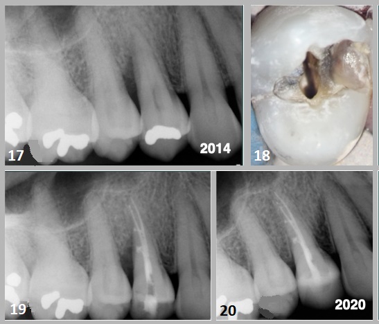
Case 4—Fig. 17: An 8mm infrabony defect used to be glaring alongside the distal proximal side of the basis of teeth #15. Fig. 18: Particles provide within the distal marginal ridge of the teeth #14 demonstrated a fracture line; alternatively, there used to be no indication of cuspal separation. Elimination of the amalgam printed extension of the fracture into the cuspal pressure aircraft of the buccal and axial line attitude. Fig. 19: The foundation canal house used to be obturated the usage of a heat vertical condensation method. Lateral branches of the basis canal gadget have been famous. Fig. 20: A post-op periapical radiograph taken in 2020 confirmed osseous regeneration and reformation of the periodontal ligament within the infrabony defect alongside the distal side of the basis.
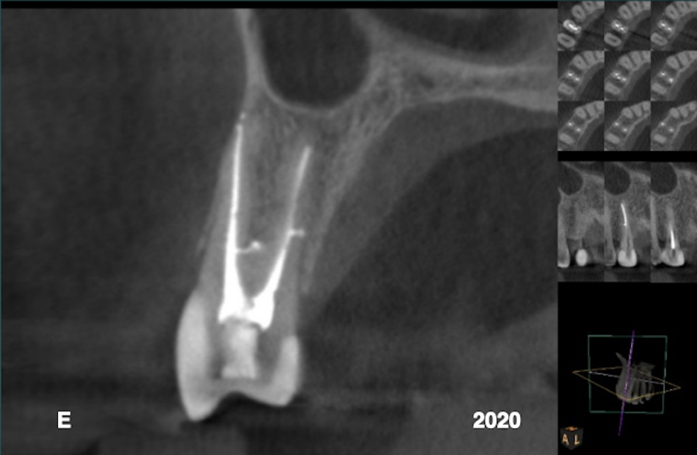
Fig. 21: A post-op periapical radiograph taken in 2020 confirmed osseous regeneration and reformation of the periodontal ligament within the infrabony defect alongside the distal side of the basis.
Conclusion
The medicine making plans procedure calls for the inclusion of a myriad of information referring to the standing of the teeth and root construction. The choice information of the American Affiliation of Endodontists encourages the clinician to believe native and systemic case-specific problems, economics, the affected person’s needs and desires, aesthetics, attainable adversarial results and moral components. The medicine carried out should mirror absolute best practices for the affected person’s wishes.
Supply By means of https://www.dental-tribune.com/information/treatment-of-compromised-teeth-the-usual-suspects/

:sharpen(level=0):output(format=jpeg)/up/dt/2023/04/Novel-coating-material-expected-to-accelerate-bone-regeneration-for-dental-implant-procedures.jpg)
:sharpen(level=0):output(format=jpeg)/up/dt/2023/04/Osstem-Implant-2022-revenue-breaks-records-in-South-Korea.jpg)